Agetic AI impact on Vascular Technology value fork gap!
Vascular Technology Ultrasound Machines using AI Machine Learning
ecto.tech
5/2/20262 min read
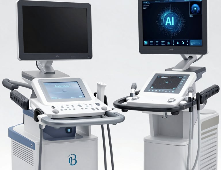
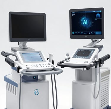
In 2026, AI and Machine Learning (ML) have shifted vascular ultrasound from a purely user-dependent diagnostic tool to an automated, predictive platform. These technologies primarily focus on reducing "operator variability"—ensuring that a scan performed by a novice looks and measures the same as one done by an expert.
Here is an elaboration on the key AI/ML features currently integrated into modern vascular ultrasound machines:
1. Automated Diagnostic Measurements
Manual measurements of vessel walls and flow are time-consuming and prone to human error. AI-driven features now handle these instantly:
AutoIMT (Intima-Media Thickness): Deep learning algorithms automatically identify the "lumen-intima" and "media-adventitia" layers of the carotid artery. It calculates the mean and maximum thickness across a segment in seconds, which is a critical marker for cardiovascular risk.
Auto-Trace Doppler: Instead of manually tracing waveforms, ML models recognize the pulse cycle and automatically calculate peak systolic velocity (PSV), end-diastolic velocity (EDV), and the resistive index (RI).
Vessel Labeling: AI can automatically identify and label anatomy (e.g., differentiating the Internal Carotid from the External Carotid) as the sonographer moves the probe.
2. Advanced Plaque Characterization
Beyond just seeing a blockage, AI can now analyze the composition of a plaque to determine how "vulnerable" or likely it is to rupture:
Plaque-IQ & Tissue Sub-typing: Software (like Elucid’s Plaque-IQ) uses ML trained on histology to quantify plaque components: calcified (stable), fibrous, or lipid-rich/necrotic core (high-risk).
Automated Stenosis Grading: AI compares the cross-sectional area of a healthy vessel segment to a diseased one to provide an exact percentage of narrowing without manual "caliper" placement.
3. Venous & Flow Dynamics
AI has recently expanded into complex venous assessments:
Venous Valve Quantification: New algorithms (like the IVENUS model) analyze "cine loops" (video) to measure the exact duration of reflux and valve opening/closing angles, which helps in grading chronic venous insufficiency.
3D Color Flow Quantification: AI assists in reconstructing 3D volumes of blood flow, providing a more accurate "Volume Flow" measurement ($mL/min$) compared to traditional 2D estimates that rely on the assumption of a perfectly circular vessel.
4. Workflow and Procedural Support
AI-Guided Needle Tracking: For vascular access (IVs or central lines), ML enhances the visualization of the needle tip and predicts its trajectory, significantly reducing "misses" and arterial punctures.
Scan Quality Feedback: Real-time AI "coaches" provide an on-screen gauge showing the quality of the image. It will tell the user to "Tilt" or "Rotate" the probe to get the optimized diagnostic view.
Autonomous Probe Positioning: Emerging robotic ultrasound systems use AI to independently find and track a vessel, allowing for hands-free monitoring during long procedures or in remote triage settings.
Summary of Benefits
Feature Traditional Method AI-Enabled Method Measurement Manual "point and click" Instant, automated contouring
Plaque Analysis Visual "guesswork" (Hypoechoic vs. Hyperechoic) Histology-based tissue quantification
Efficiency High number of keystrokes ~50% reduction in exam time
Consistency Highly operator-dependent Standardized results across users
Results
Experienced team delivering exceptional results for your business.
Gap-Connection
Marketing
© 2024. All rights reserved.